Types of Cataract Surgery – Overview
Cataract surgery, also known as lens replacement surgery is the surgical removal of the natural lens of the eye and replacement with an artificial intraocular lens.
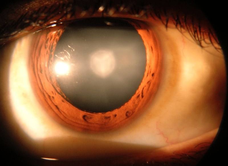
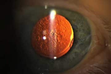
The procedure is carried out to treat cataracts, a gradual clouding of the eye lens which leads to a decrease in vision.
Symptoms of cataracts may include faded colors, blurry or double vision, halos around light, trouble with bright lights, and trouble seeing at night.
This may result in trouble driving, reading, or recognizing faces. Poor vision caused by cataracts may also result in an increased risk of falling and depression. Cataracts cause half of all cases of blindness and 33% of visual impairment worldwide.
Cataract surgery is performed to treat the medical condition and there are different types of cataract surgery.

Types of Cataract Surgery
There are three main types of cataract surgery namely; Phacoemulsification, Extracapsular cataract surgery, Intracapsular cataract surgery.
1. Phacoemulsification
Also known as ‘Phaco’, it is the most common technique used for cataract removal nowadays.
Usually, it takes no more than half an hour to remove cataracts through phacoemulsification, and that too, requires only minimal sedation, i.e. local anesthesia (injecting anesthesia around the eye) or topical anesthesia (administering numbing drops into the eye).
This cataract procedure requires a small surgical incision around the edge of the cornea, creating an opening through the membrane surrounding the lens. The next step involves inserting a small ultrasonic probe into the opening to break up the cloudy lens into tiny fragments using sound waves, which act as a microscopic jackhammer. An attachment on the probe tip is then used for suction of broken-down cataract fragments.
Once the lens particles are removed, an intraocular lens implant, also referred commonly to as an IOL, is implanted in the natural lens capsule. A hollowed-out tube is used by ophthalmologists to insert the IOL through a tiny corneal incision.
2. Extracapsular cataract surgery
This is the cataract procedure used in the case of highly advanced cataracts, which is too dense for phacoemulsification (the process of breaking down or dissolving the cataract into tiny fragments) or when phacoemulsification is not possible for various other reasons.
A slightly larger incision is required for this cataract removing technique so that the cataract can be removed in one piece instead of being fragmented within the eye. Just like phacoemulsification, an artificial lens (IOL) is placed inside the same capsular bag.
Several sutures are required to close the comparatively larger wound, which also results in slower recovery of the wound as well as visual function.
To initiate this cataract removal technique, the numbing medication is administered through an injection around the eye. An eye patch is also required after this kind of surgical process.
3. Intracapsular cataract surgery
Though rarely used nowadays, this cataract removal technique may still be useful under certain circumstances. It requires an even larger incision as compared to extracapsular surgery, through which the entire lens with surrounding capsule is removed. Moreover, the IOL (intraocular lens) is placed in a different location, in front of the iris, in this surgical procedure.
Surgical Procedure
Cataract surgery is performed by an ophthalmologist on an outpatient basis, i.e no hospital stay is required. There is the application of local anesthesia to the eyes which prevents the patient from feeling any pain while remaining awake.
The procedure requires only very small incisions, and no stitches are needed at the end of surgery. Hand-held instruments are used to gain access to the cataract, which is then broken up with ultrasonic energy and removed from the eye in small fragments with gentle suction.
A special applicator is then used to insert the flexible IOL, which unfolds inside the eye. The surgeon securely positions the IOL in the same location where the natural lens resided (directly behind the pupil).
Related: Eye Muscle Surgery
Risks and Complications
In as much as cataract surgery has a high success rate of about 95%, there is still the risk of complications as in any surgical procedure. Some possible complications with cataract surgery include:
- Infection – Endophthalmitis is a serious infection of the intraocular tissues, usually following intraocular surgery, or penetrating trauma. There is some concern that the clear cornea incision might predispose to the increase of endophthalmitis but there is no conclusive study to corroborate this suspicion.
- Bleeding – There may be excessive bleeding during or after the surgery
- Retina detachment – This is a disorder of the eye in which the retina separates from the layer underneath. The prevalence rate of complications following cataract surgery is about 1 in 1000.
Other possible complications include Swelling or edema of the cornea, sometimes associated with cloudy vision, which may be transient or permanent (pseudophakic bullous keratopathy). Displacement or dislocation of the intraocular lens implant may rarely occur.
Unplanned high refractive error (either myopic or hypermetropic) may occur due to error in the ultrasonic biometry (measure of the length and the required intraocular lens power). Cyanopsia, in which the patient sees everything tinted with blue, often occurs for a few days, weeks, or months after the removal of a cataract. Floaters commonly appear after surgery.
Cataract Surgery Cost
The total cost for cataract surgery depends on a lot of factors such as the anesthetic fee, private hospital fee, private operating facility fee, type of surgery required. The total cost of the procedure is around $3,500 – $7000. Medicare covers cataract surgery if you don’t have health insurance.
Cataract Recovery
Cataract surgery recovery is also one of the most concerning aspects of cataract removal, especially the time required for restoration of functional vision after the procedure.
The patient is expected to take the first follow-up visit to their ophthalmologist within the first few days after the cataract surgery, followed by a subsequent visit within the first few weeks. In most cases, there is the prescription of specific types of eye drops to reduce inflammation and protect against infection.
Most people observe an improvement in their visual function within several days of the surgery, enabling them to restart most of their daily activities including work.
However, the patient needs to have several visits to the doctor, who monitors and observes the eye(s) for any signs of complications.
The majority of people regain full activity after a cataract recovery time of a few days of getting treated. Your doctor might fit you with a pair of glasses (if needed) once your vision is stabilized. This also depends upon the type of intraocular lens implanted.
More: Types of Hip Surgery


