Types of Hip Fractures
Overview
A hip fracture is a break in the femur’s upper part (thigh bone). The majority of hip fractures happen in older people who already have osteoporosis, which has damaged their bones. A high-energy accident, including a falling from a height or a car crash, is usually the cause of a hip fracture in a younger patient.
A hip fracture affects over 270,000 individuals in the USA each year. Patients 60 years and older who are injured in the house or in public accidents account for the majority of these fractures.
Hip fractures are excruciatingly painful. As a result, early surgical intervention is advised.
Medical consequences such as pressure sores, deep vein thrombosis (obstruction that occurs in leg veins), and pneumonia (chest infection) should be prevented by treating the fracture and bringing the patient out of bed as soon as feasible.
Prolonged bed rest can confuse elderly individuals, making physiotherapy and recovery extra challenging.
Structures of Hip Affected by Fractures

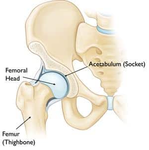
Types of Hip Fractures – Structure of the Hip
Inter-trochanter. The space between the femur’s neck and the femur’s shaft (the long portion of the femur). The greater trochanter and lesser trochanter are two bony structures that define it as intertrochanteric.
The neck of the femur. the femur’s portion below the femur’s head.
With inter-trochanter area, they are the most commonly fractured structures in the hip joint
Sub-trochanteric. The higher portion of the femur’s shaft is below both trochanters.
Head of the femur. The femur’s ball fits in the hole of the pelvic bone.
Symptoms of Hip Fractures
• Pain
• Having difficulty raising, moving, or turning your limb
• Being unable to erect or bear weight on one’s leg
• Bruises and puffiness in the region of your hip
• The affected limb appears to be shorter than the other.
• Curving outwards of your affected leg
A hip fracture does not always result in bruises or the inability to stand or move.
Causes of hip fractures
• Falling from a considerable height or onto a hard surface.
• An automobile accident, for example, can cause massive trauma to the hip.
• In older people with weak bones, fractures can occur simply while walking.
Risk Factors of Hip Fractures
• Being thin.
• Deficiency in calcium or vitamin D.
• Osteoporosis (bone loss)
• Physical inactivity
• Consuming an excessive amount of alcohol
• Smoking
Diagnosis of Hip Fractures
In addition to a comprehensive medical history and clinical assessment, diagnostic techniques for hip fractures may involve the following:
• X-ray. Internal tissues, bones, and organs are captured on film by invisible electromagnetic radiation waves.
• MRI. Powerful magnets, radio waves, and a computer work together to create high-resolution images of your organs and structures.
• CT scan. This is an imaging modality that makes high-resolution images of the body using X-rays and a computer. The bones, muscles, fat, and organs are all visible on Computed tomography. CT scans provide further information than traditional X-rays.
Intertrochanteric Fracture
Extracapsular fractures of the proximal femur that occur between the greater and lesser trochanters are known as intertrochanteric fractures.
The intertrochanteric component of the femur is made up of compact trabecular bone that lies between both the greater and lesser trochanters.
An intramedullary nail or a gliding compressive hip screw and lateral plate are used to fix intertrochanteric fractures surgically.
Bone screws secure the compressive hip screw to the exterior surface of the femur. A large screw (lagging screw) is inserted into the femoral head and neck via the plate.
The impaction and compression at the fractured site are made possible by this arrangement. This will improve stability and aid in the healing process.
Through an incision at the apex of the greater trochanter, the intramedullary nail is directly integrated into the medullary canal of the bone. After that, one or more screws are driven through the nail and into the femoral head.
Femoral Neck Fracture

Types of Hip Fractures
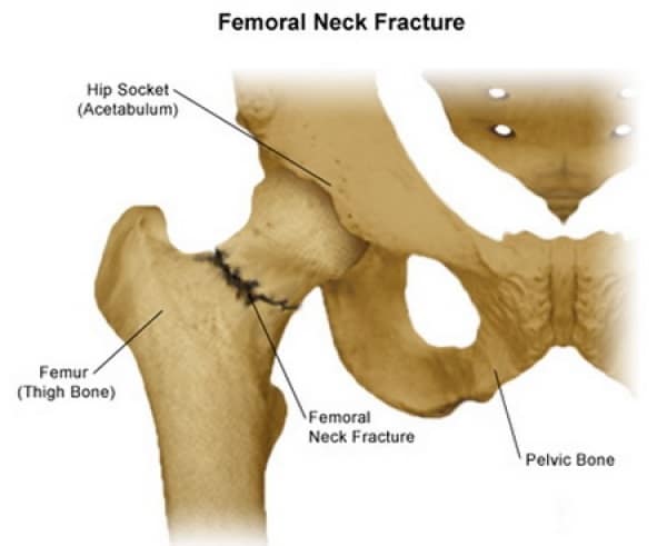
A femoral neck fracture occurs when the femur is fractured immediately below the head of the roller bearings hip joint. The head of the femur is separated from the remainder of the femur in this form of fracture. Groin pain is common, and it gets worse when you put weight on the afflicted leg.
pinning is the most popular method for a neck or femur fracture that is not dislocated. Surgical rods or screws are inserted all across the fractured site to keep the femur’s head in place as it heals.
The femoral head is pinned to keep it from detaching or sliding off the femoral neck, which would necessitate joint replacement.
due to the risk of a condition called femoral neck necrosis, Hip replacement is widely used to treat dislocated femoral neck fractures. partial hip replacement is the treatment option of choice for older patients. Total hip replacement may be recommended in younger, more active patients.
Sub-trochanteric Fracture
The upper portion of the femoral shaft, right below the hip joint, is damaged by a sub-trochanteric fracture.
These fractures are surgically repaired by inserting an intramedullary nail into the proximal femur and a screw into the femoral head through the nail.
Additional screws may be put at the lower end of the nail at the knee to prevent the bones from twisting around the nail or shortening the nail.
In some circumstances, instead of a nail, your surgeon may choose a compression screw with a lengthy side plate.
Femoral Head Fracture
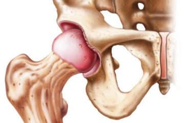
Femoral head fractures are uncommon; only around 1% of all hip fractures are femoral head fractures. They are frequently the outcome of a high-velocity incident. There is a chance that the hip joint socket will be fractured as well.
Nonsurgical treatment with limited weight-bearing is possible if the fracture is not displaced. If the displaced fragment is minor and does not cover a considerable portion of the joint surface, it can be easily removed.
When a big fragment is present in a young and active person, open reduction and internal fixation are frequently used. Hip replacement—partial or total—to replace the injured femoral head is the treatment of choice for an elderly patient.
See Also
Hip Joint Surgery
Postural Orthostatic Tachycardia Syndrome
Types of Surgery
References
https://www.mayoclinic.org/diseases-conditions/
https://www.nhs.uk/conditions/hip-fracture/
https://www.cedars-sinai.org/health-library/

Dr.Sharif Samir Alijla, is a general medical doctor and a well-rounded professional that cares and treats patients from Palestine. I participated in many medical studies and conferences, I've launched a range of community initiatives and taken part in a variety of leadership and change training programs. I worked as an author for many medical websites such as TebFact . I specialized in writing medical articles from authoritative and updated sources in a simple and smooth the way for the reader.


